Home » Without Label » Foot Muscles Mri - The MRI Technologist Role in Foot/Ankle Positioning | All ... - Mri with user outlined plantar intrinsic and extrinsic muscles group.
Foot Muscles Mri - The MRI Technologist Role in Foot/Ankle Positioning | All ... - Mri with user outlined plantar intrinsic and extrinsic muscles group.
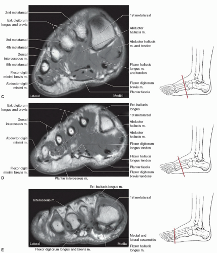
Foot Muscles Mri - The MRI Technologist Role in Foot/Ankle Positioning | All ... - Mri with user outlined plantar intrinsic and extrinsic muscles group.. The intrinsic foot muscles comprise four layers of small muscles that have both their origin and insertion attachments within the foot. Ultrasonography seems to be useful for detection of foot muscle atrophy in diabetes. Those fibers of the most medial and largest belly are known as. A magnetic resonance imaging (mri) was performed on a normal subject; Several authors have hypothesised that intrinsic muscle weakness is an important contributor to the development of pes cavus deformity ,.
Muscle anatomy labeling 12 photos of the muscle anatomy labeling anatomy muscle labeling games, holes anatomy muscle labeling, mcgraw hill anatomy muscle labeling, muscle anatomy model labeled, skeletal muscle anatomy labeling, human muscles, anatomy muscle labeling games, holes anatomy muscle labeling, mcgraw hill anatomy. The aim of this review is to provide the reader with a comprehensive overview of the magnetic resonance imaging (mri) characteristics of the most common benign and malignant soft tissue neoplasms which occur around the foot and ankle. Magnetic resonance imaging (mri) is the modality of choice in diagnosing accessory muscles, delineating their relationship to adjacent structures, and differentiating them from soft tissue tumors. T he complex anatomy of the foot and ankle makes imaging of this region challenging. Adductor hallucis is anatomically located in the central compartment of foot, but the muscle is functionally grouped with the medial plantar muscles of foot because it acts on the great toe (hallux).
Foot, Ankle, and Calf | Musculoskeletal Key from musculoskeletalkey.com Magnetic resonance imaging of foot and ankle pathology. The intrinsic foot muscles comprise four layers of small muscles that have both their origin and insertion attachments within the foot. Accessory muscles are isointense to skeletal muscle on all pulse sequences, and can insert by fleshy muscular or tendinous insertions. Editor · aug 14, 2017 ·. The aim of this review is to provide the reader with a comprehensive overview of the magnetic resonance imaging (mri) characteristics of the most common benign and malignant soft tissue neoplasms which occur around the foot and ankle. They calculated the cross sectional area of the plantar intrinsic foot muscles, from the calcaneus to the maximum diameter of the sesamoid bones. Hip pelvis thigh knee lower extremity/shin ankle foot. Coronal images are perpendicular to the long axis of the metatarsals.
Spine bone tumor/mets yes mri cervical, thoracic or lumbar with and without discitis yes mri cervical, thoracic or lumbar with and without extremity numbness/tingling no mri cervical, thoracic or lumbar without history of lumbar surgery yes mri lumbar with and without multiple sclerosis yes mri cervical or thoracic with and without
Accessory muscles are isointense to skeletal muscle on all pulse sequences, and can insert by fleshy muscular or tendinous insertions. Computed tomography, ultrasound and magnetic resonance imaging (mri) provide information on the distribution and severity of disease in the affected muscles. T he complex anatomy of the foot and ankle makes imaging of this region challenging. Related posts of foot muscle anatomy mri muscle anatomy labeling. A magnetic resonance imaging (mri) was performed on a normal subject; These findings are important and relevant for clinicians involved in the assessment and treatment of foot and lower. The muscles acting on the foot can be divided into two distinct groups; Causative lesions may be apparent such as: The aim of this review is to provide the reader with a comprehensive overview of the magnetic resonance imaging (mri) characteristics of the most common benign and malignant soft tissue neoplasms which occur around the foot and ankle. Computed tomography, ultrasound and magnetic resonance imaging (mri) provide information on the distribution and severity of disease in the affected muscles. Foot muscles mri anatomy / plantar tendons of the foot mr imaging and us radiographics / neuropathies around the elbow joint. A sagittal image of a foot representing the localization of serial axial mri (a).a typical example of mri with a manually painted three plantar intrinsic muscle groups (b).a sagittal image of a lower leg representing the localization of serial axial mr images (c).a typical example of the analyzed image for two plantar. Near normal foot mri for reference.
Mri with user outlined plantar intrinsic and extrinsic muscles group. Editor · aug 14, 2017 ·. T he complex anatomy of the foot and ankle makes imaging of this region challenging. The adductor hallucis has two heads: Magnetic resonance imaging (mri), with its multiplanar capabilities, superior soft tissue contrast, excellent spatial resolution, ability to image bone marrow, noninvasiveness, and lack of.
MRI Protocols: ANKLE MRI IMAGING PLANES from lh6.googleusercontent.com Magnetic resonance imaging of foot and ankle pathology. The muscular system is responsible for the movement of the human body. Muscle anatomy labeling 12 photos of the muscle anatomy labeling anatomy muscle labeling games, holes anatomy muscle labeling, mcgraw hill anatomy muscle labeling, muscle anatomy model labeled, skeletal muscle anatomy labeling, human muscles, anatomy muscle labeling games, holes anatomy muscle labeling, mcgraw hill anatomy. They are mainly responsible for assisting some of the extrinsic muscles in their actions. Related posts of foot muscle anatomy mri muscle anatomy labeling. Chang and colleagues analyzed the feet of eight subjects with unilateral plantar fasciitis, using a 1.5 tesla magnetic resonance imaging system. Weakness of intrinsic foot muscles is a widely accepted pathological finding of cmt and magnetic resonance imaging (mri) studies have indicated significant atrophy in intrinsic foot muscles ,. Magnetic resonance imaging of musculoskeletal soft tissue masses.
Weakness of intrinsic foot muscles is a widely accepted pathological finding of cmt and magnetic resonance imaging (mri) studies have indicated significant atrophy in intrinsic foot muscles ,.
Both muscles are innervated by the deep fibular nerve. Several authors have hypothesised that intrinsic muscle weakness is an important contributor to the development of pes cavus deformity ,. Atrophy of intrinsic foot muscles determined at ultrasonography is directly related to foot muscle volume determined by mri and to various measures of diabetic neuropathy. Adnan sheikh, marcos loreto sampaio and mark e. Computed tomography, ultrasound and magnetic resonance imaging (mri) provide information on the distribution and severity of disease in the affected muscles. This imaging technique assesses the ligaments and tendons, neurovascular structures (tarsal tunnel and plantar fascia), and the osseous structures(19). In the very early stage, muscle signal may be normal. Other important tendons in the foot include the tibialis posterior (posterior tibial tendon), which attaches the calf muscle to the bones on the inside of the foot and supports the arch of the foot, and the tibialis anterior (anterior tibial tendon), which runs from the outer tibia to the first metatarsal and surfaces of the median cuneiform tarsal, which allows for dorsiflexion—bringing the. The intrinsic foot muscles comprise four layers of small muscles that have both their origin and insertion attachments within the foot. Mri is the choice of modality for further imaging the ankle and foot after obtaining initial radiographs. • muscle edema is seen secondary to multiple etiologies including trauma, infectious and inflammatory processes, autoimmune disorders, neoplasms, and denervation injuries • on mri muscle edema is characterized by increase in free water within the muscle • muscle edema is seen on mri as increased signal on fluid sensitive sequences t2 fs As the fiber bundles extend distally, they become grouped into four bellies. Anatomy of the whole human body :
T he complex anatomy of the foot and ankle makes imaging of this region challenging. The muscular system is responsible for the movement of the human body. Chang and colleagues analyzed the feet of eight subjects with unilateral plantar fasciitis, using a 1.5 tesla magnetic resonance imaging system. Muscle anatomy labeling 12 photos of the muscle anatomy labeling anatomy muscle labeling games, holes anatomy muscle labeling, mcgraw hill anatomy muscle labeling, muscle anatomy model labeled, skeletal muscle anatomy labeling, human muscles, anatomy muscle labeling games, holes anatomy muscle labeling, mcgraw hill anatomy. In the very early stage, muscle signal may be normal.
MRI foot muscles - RadiologyER from 1.bp.blogspot.com Effects of direct injury or tear. Adnan sheikh, marcos loreto sampaio and mark e. Hip pelvis thigh knee lower extremity/shin ankle foot. Distal part of the lateral and superior surfaces of the calcaneus and the apex of the inferior extensor retinaculum. In addition, an image of all the muscles of the back and. Anatomy of the whole human body : Foot muscles mri anatomy / plantar tendons of the foot mr imaging and us radiographics / neuropathies around the elbow joint. Thank you for your attention.
Near normal foot mri for reference.
Muscle anatomy labeling 12 photos of the muscle anatomy labeling anatomy muscle labeling games, holes anatomy muscle labeling, mcgraw hill anatomy muscle labeling, muscle anatomy model labeled, skeletal muscle anatomy labeling, human muscles, anatomy muscle labeling games, holes anatomy muscle labeling, mcgraw hill anatomy. This is a 30 year old with swelling on the lateral aspect of foot with evidence of soft tissue lesion in relation to the lateral aspect of the talus which appears isointense to the muscles on t1 and t2 weighted images & appears elongated extending from the anterosuperior calcaneum to the base of the 5th metatarsal. Magnetic resonance imaging of foot and ankle pathology. Both muscles are innervated by the deep fibular nerve. When the muscles tighten (contract) they pull on the tendons, which in turn move the bones. Mri of the soft tissues of the foot visualizes the fat cushions of the sole, heels, fingers and can show swelling, foci of. • muscle edema is seen secondary to multiple etiologies including trauma, infectious and inflammatory processes, autoimmune disorders, neoplasms, and denervation injuries • on mri muscle edema is characterized by increase in free water within the muscle • muscle edema is seen on mri as increased signal on fluid sensitive sequences t2 fs In the very early stage, muscle signal may be normal. Causative lesions may be apparent such as: Magnetic resonance imaging (mri) is the modality of choice in diagnosing accessory muscles, delineating their relationship to adjacent structures, and differentiating them from soft tissue tumors. Ultrasonography seems to be useful for detection of foot muscle atrophy in diabetes. Chang and colleagues analyzed the feet of eight subjects with unilateral plantar fasciitis, using a 1.5 tesla magnetic resonance imaging system. Those fibers of the most medial and largest belly are known as.